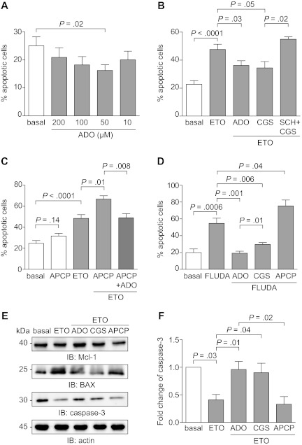
Figure 7.
Extracellular ADO protects from both spontaneous and drug-induced apoptosis. (A) Purified CLL cells were plated with or without extracellular ADO at the indicated doses. The percentage of apoptotic cells was determined after 16 hours. n = 10. (B) CLL cells were cultured in the presence of etoposide (50μM) in combination with ADO (50μM). Where indicated, CLL cells were pretreated with CGS21680 (10μM, for 30 minutes at 37°C) alone or in combination with SCH58261 (10μM, for 30 minutes at 37°C). n = 10. (C) CD73 enzymatic activity was blocked using the APCP inhibitor (10μM, for 60 minutes at 37°C). Pretreatment of CLL cells with APCP in the presence of etoposide (50μM) significantly increase the apoptotic rate, corrected by the addition of exogenous ADO (50μM). n = 10. (D) Purified CLL cells were treated with fludarabine (5μM), alone or in combination with ADO (50μM), and apoptosis was evaluated after 48 hours. Where indicated, CLL cells were pretreated with CGS21680 or APCP (10μM, for 30 minutes and 60 minutes at 37°C, respectively). n = 10. (E) Western blot analysis of the expression of the p53-dependent genes Mcl-1, BAX, and caspase-3, after the exposure of CLL cells to etoposide (50μM) as DNA-damaging agent. (F) Quantitative analysis of caspase-3 expression. Fold change is represented as the ratio of each treatment on the basal condition. n = 5. Error bars represent the SEM in all the graphs. Statistical analysis was performed using Student t or Wilcoxon signed rank tests. SCH indicates SCH58261; ETO, etoposide; and FLUDA, fludarabine.

