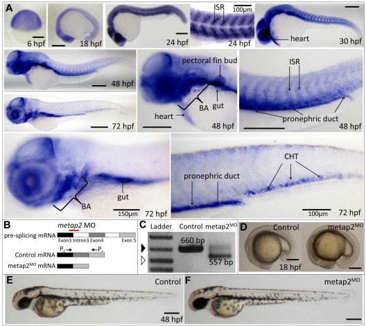
Figure 1.
Expression of metap2 during embryonic development and metap2 knockdown with morpholino. (A) WISH showing the spatial expression of metap2 at different developmental stages. BA indicates brachial arches; ISR, intersomitic region. (B) Diagram of the action of the metap2 morpholino. Red line represents the binding site of the splice-junction targeted metap2 morpholino, and arrows represent the binding sites of the forward (Pf) and reverse primers (Pr) used for PCR amplification to detect defective splicing of exon4. (C) Defective splicing of metap2 mRNA in metap2MO as shown by the presence of the smaller 557-bp band, which is absent in the control. Open arrowhead, 500-bp DNA ladder; filled arrowhead, 650-bp DNA ladder. (D) General morphology of metap2MO (122 of 131 embryos in 3 separate experiments) compared with the control at 18 hpf (137/145, n = 3). (E-F) General morphology of metap2MO (F; 109/129, n = 3) compared with the control (E; 132/142, n = 3) at 48 hpf. Bars represent 250 μm unless otherwise stated.

