Figure 3.
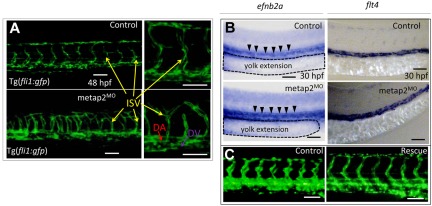
Knockdown of metap2 perturbed angiogenesis but not vasculagenesis. (A) Confocal microscopy of Tg(fil1:gfp) at 48 hpf comparing the development of dorsal aorta (DA), dorsal vein (DV), and intersegmental vessel (ISV) in the trunk region between control and metap2MO. (B) WISH comparing expression of efnb2a (control, 92.4% ± 2.3% with normal efnb2a expression in total 92 embryos; metap2MO, 91.4% ± 2.7% with normal efnb2a expression in total 93 embryos; n = 3) and flt4 (control, 95.5% ± 1.4% with normal flt4 expression in total 88 embryos; metap2MO: 95.3% ± 1.0% with normal flt4 expression in total 86 embryos; n = 3) at 30 hpf between control and metap2MO. Black arrowheads denote efnb2a expression along DA, and the dashed line outlines the extension of the yolk. (C) Fluorescent microscopy of Tg(fil1:gfp) at 48 hpf comparing the development of DA, DV, and ISV in the trunk region between control and metap2MO rescued by metap2 mRNA (control, 96.8% ± 1.4% with normal ISV patterning in total 93 embryos; metap2MO, 80.3% ± 3.4% with perturbed ISV patterning in total 71 embryos; metap2MO rescue, 72.5% ± 4.0% with normal ISV patterning in total 80 embryos; n = 3). Bars represent 50 μm.

