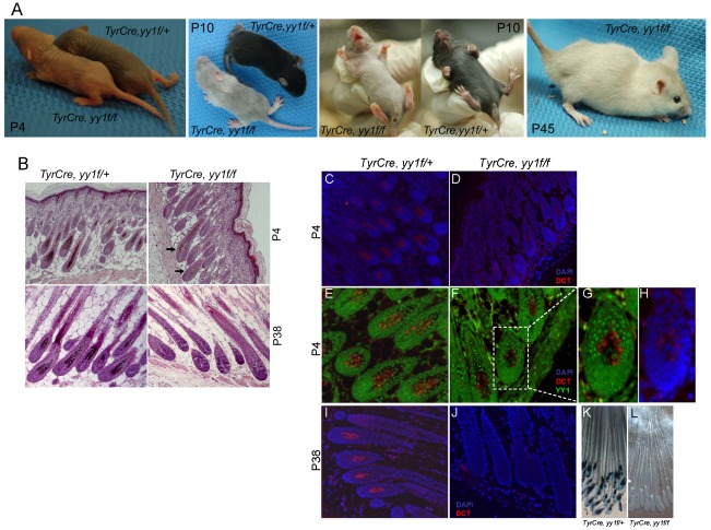
Figure 1. YY1 is required for melanocyte development in vivo.
(A) Skin and hair pigmentation phenotype of TyrCre, yy1f/fl in the first (P4, P10) and second (P45) hair cycles. (B) H&E staining of hair follicles from skin sections of TyrCre, yy1f/+ and TyrCre, yy1f/f mice in the first (P4) and second (P38) hair cycles. Arrows point to the hair follicles still containing pigment. (C,D) Immunofluorescence staining of DCT (red) in P4 hair follicles of TyrCre, yy1f/+ (C) and TyrCre, yy1f/f (D) mice. Skin sections were stained with goat anti-DCT primary antibody and donkey anti-goat Alexa 594 secondary antibody. Nuclei were counterstained with DAPI (blue). (E,F) Immunofluorescence staining of DCT (red) and YY1 (green) in P4 hair follicles of TyrCre, yy1f/+ (E) and TyrCre, yy1f/f (F) mice. Skin sections were double-stained with goat anti-DCT and rabbit anti-YY1 primary antibodies and then donkey anti-goat Alexa 594 and goat anti-rabbit Alexa 488 secondary antibodies. (G,H) Zoom-in view of the dashed box area in (F). DAPI stain is in blue in (H). (I,J) Immunofluorescence staining of DCT (red) in P38 hair follicles of TyrCre, yy1f/+ (I) and TyrCre, yy1f/f (J) mice. Nuclei were stained with DAPI (blue). (K&L) XGal stain of whole-mount skin sections from P38 TyrCre, yy1f/+, Dct-LacZ (K) and TyrCre, yy1f/f, Dct-LacZ (L) mice.

