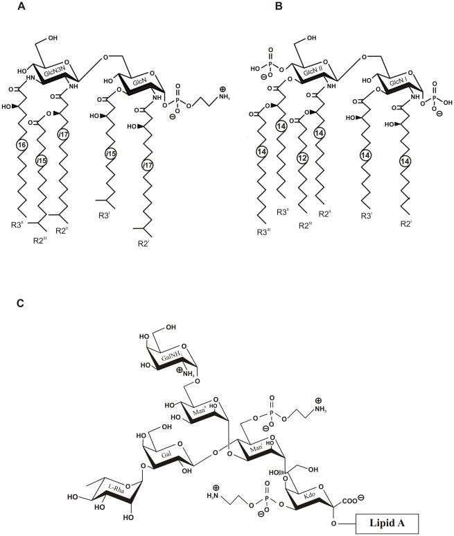
Figure 2. Structures of C. canimorsus lipid A, E. coli lipid A and core-oligosaccharide of C. canimorsus attached to the lipid A.
(A) C. canimorsus lipid A consists of a β-(1′→6)-linked GlcN3N′-GlcN disaccharide, to which 3-hydroxy-15-methylhexadecanoic acid, 3-hydroxy-13-methyltetradecanoic acid, 3-O-(13-methyltetradecanoyl)-15-methylhexadecanoic acid, and 3-hydroxyhexadecanoic acid are attached at positions 2, 3, 2′, and 3′, respectively. The disscharide carries a positively charged ethanolamine at the 1 phosphate and lacks a 4′ phosphate. (B) Structure of E. coli hexa-acylated lipid A. (C) C. canimorsus LPS core features only one Kdo, to which a phosphoethanolamine (P-Etn) is attached. The only net negative charge present is from the carboxy group of the Kdo. The inner core continues with Man to which another a P-Etn is attached. The outer core consists of Gal and l-Rhamnose (l-Rha), to which the O-antigen is attached (U. Zähringer, unpublished results).

