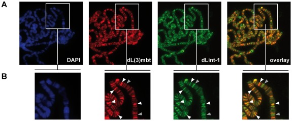
Figure 3. dL(3)mbt and dLint-1 colocalise on polytene chromosomes.
(A) Immunofluorescence stainings of polytene chromosomes. Flies carrying an dL(3)mbt transgene under control of UAS were crossed with a salivary gland-specific sgs58AB-GAL4 driver strain. Polytene chromosomes were stained with dL(3)mbt, dLint-1 antibodies and DAPI as indicated. The right panel shows an overlay of the dL(3)mbt and dLint-1 staining. (B) Magnified sections of the panels shown in (A). White and grey arrow heads denote selected prominent sites of colocalization (white) and exclusive dLint-1 binding sites (grey), respectively.

