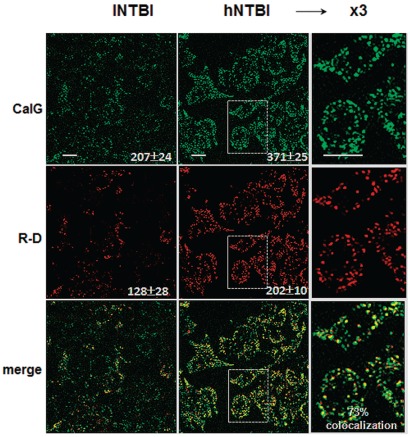
Figure 4.
Endocytosis of CALG and rhodamine-dextran by RAW cells in media containing sera from thalassemia major patients. RAW cells were incubated for 3 h with thalassemia major sera containing low and high NTBI (lNTBI or hNTBI) as indicated and both CALG (10 μM) and Rhodamine-dextran (R-D) (30 μM). After washing of cells and treatment with SIH 50 μM, the fluorescence of CALG and R-D was monitored by live epifluorescence microscopy equipped with an Optigrid system. The fluorescence intensity of CALG and R-D obtained from 5 different cell areas and the mean values obtained from 3 independent experiments are shown in the body of the figure. Colocalization of merged images of CALG and R-D (shown in the 3rd row of images) performed with the Volocity program was 73–75%.

