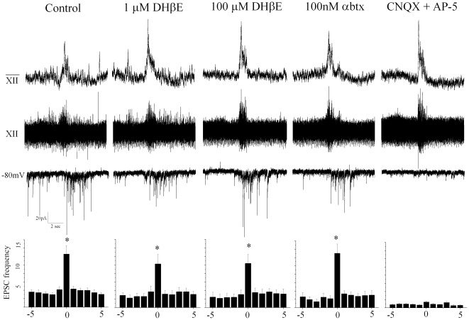
Figure 2. Respiratory-related GABAergic neurons receive glutamatergic input that is blocked by antagonists CNQX and AP-5.
Glutamatergic receptors mediate excitatory neurotransmission to inspiratory GABAergic neurons. Inspiratory-related bursting activity was recorded from the hypoglossal rootlet (top trace) and electronically integrated (middle trace). Excitatory transmission to GABAergic neurons (bottom trace) was isolated by focal application of GABA (gabazine; 25 µM) and glycine (strychnine; 1 µM) receptor antagonists. Under control conditions there was a significant increase in EPSC frequency in the identified GABAergic neurons during inspiratory bursts control spontaneous: 3.5+/−0.5, control inspiratory: 12.6+/−1.9 Hz. DHßE (at concentrations of 1 µM, n = 8 and 100 µM, n = 7), and α-Btx (100 nM, n = 6) did not significantly change spontaneous or inspiratory related EPSC frequency. CNQX and AP5 blocked EPSC spontaneous and inspiratory frequency (p<0.05). The average results from all neurons tested (DHßE at 1 µM n = 8, at 100 µM n = 7, and with α-Btx at 100 nM, n = 6) are shown in the peri-inspiratory activity histogram, bottom, illustrating the average EPSC frequency (Hz) for each of the 5 seconds before the burst, frequency during the inspiratory burst (which typically lasted 1–2 seconds), and each of the 5 seconds following the inspiratory burst.

