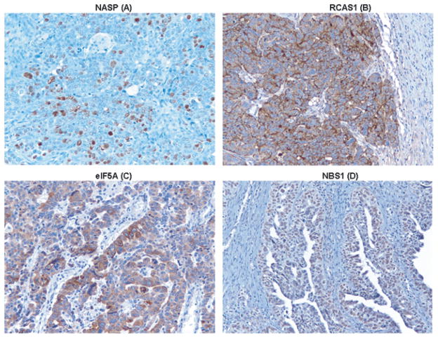
Fig. 1.
Immunohistochemical staining for NASP, RCAS1, eIF5A and NBS1 in tissue sections obtained from a patient with serous high grade carcinoma. IHC was performed to determine the protein expression level of 4 serological biomarkers (mentioned above) that were previously identified by profiling humoral immune responses in OVCA patients [6]. Nuclear staining was observed for NASP (A), cytoplasmic staining was observed for RCAS1 (B) and eIF5A (C), nuclear staining was observed for NBS1 (D). Magnification in all panels is X20.

