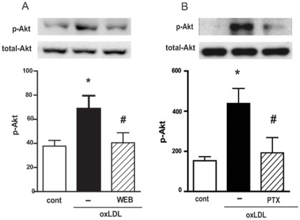
Figure 1. oxLDL activates the PI3k/Akt pathway in a PAFR and Gαi coupled protein-dependent manner.
Adherent monocytes/macrophages were treated with WEB2170 (50 µM, 30 min) (A) or PTX (600 ng/mL, 18 h) (B) and stimulated with oxLDL (30 µg/mL) for 10 min. Western blot analysis of cell lysates were performed using antibodies to phosphorylated and non-phosphorylated forms of Akt. Data are presented as mean ± SEM of four donors. Protein expression was quantified by the AlphaEaseFC™ software V3.2 beta (Alpha Innotech). The autoradiographs show one representative experiment. * p<0.05 comparing oxLDL-stimulated with the non-stimulated cells (cont). # p<0.05 comparing cells treated with WEB2170 or PTX with non-treated cells.

