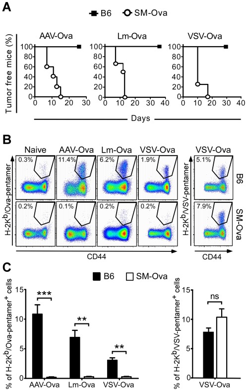
Figure 1. Lack of detectable Ova-specific CD8+ T cell responses in immunized SM-Ova mice.
(A) B6 or SM-Ova mice were immunized with rAAV-Ova, replicative VSV-Ova or live Lm-Ova. Seven days after infection with VSV-Ova or Lm-Ova, or 14 days after injection of rAAV-Ova, mice were inoculated with 1×10⁶ Ova-bearing EG-7 tumor cells and monitored during 30–40 days for tumor development. (B) In other groups of mice, animals were immunized with the same vaccines and splenocytes were analyzed 7 or 14 days after by flow cytometry to evaluate the percentage of CD8+ T cell recognizing the immunodominant peptides of Ova or VSV-associated nucleoprotein using H-2Kb/Ova257–264 or H-2Kb/VSV pentamers staining, respectively. Representative flow cytometry profiles are shown and numbers indicate percentage of pentamer-positive cells in the CD8+-gated population. Background staining using H-2Kb/VSV pentamers were always below 0.3% of CD8+ cells as also shown for the staining using H-2Kb/Ova257–264 pentamers (C) Bar graphs represent mean percentages of CD8+ T cells positively stained with the indicated H-2Kb/Ova257–264 or H-2Kb/VSV pentamers in B6 (black bars) or SM-Ova (open bars). Data are representative of 3 independent experiments, each one performed with 5–7 mice per group.

