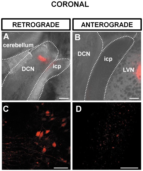
Figure 2. Coronal brainstem slice showing a retrograde labelling of LVN following injection of dextran amine in the DCN (A,C) and an anterograde labelling of the DCN following injection of dextran amine in the LVN (B,D).
(A) Overlay of a brightfield and fluorescence photomicrograph at 3 hours post injection of dextran amine showing the position of the DCN relatively to the inferior cerebellar peduncle (icp) and the cerebellum. The fluorescence in the DCN shows the injection site. (B) The LVN is labeled as a result of retrograde transport of dextran amine. (C) Overlay of a brightfield and fluorescence photomicrograph showing the position of the dextran amine injection site in the LVN. (D) Labeled terminals in the DCN as a result of anterograde transport of dextran amine. Scale bar: (A) and (B) 200 µm, (C) and (D) 20 µm. All slices are 120 µm thick.

