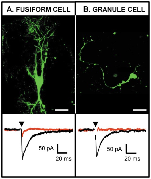
Figure 4. Glutamatergic post-synaptic currents (EPSCs) elicited in identified DCN cells by stimulating the LVN in a sagittal slice.
(A) Photomicrograph of a DCN fusiform cell filled with lucifer yellow (top) and whole cell voltage clamp recording of this fusiform cell while stimulating the LVN (bottom). (B) Photomicrograph of a DCN granule cell filled with lucifer yellow (top) and whole cell voltage clamp recording of this granule cell while stimulating the LVN (bottom). Both cells were held at −68 mV and the LVN was stimulated at 0.3 Hz. Glutamatergic EPSCs are represented in black and are blocked by 50 µm D-AP5 and 10 µm NBQX (traces in red). Each trace represents an average of 10–20 single traces. The arrowhead represents the artifact of stimulus that has been removed for clarity. Scale bar: (A) 50 µm, (B) 20 µm.

