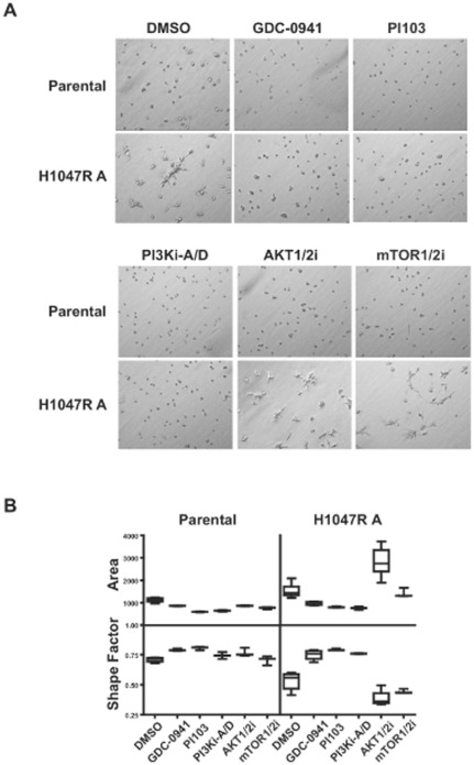
Figure 3. MCF10A knock-in cells show a more invasive phenotype in 3-D cell culture.
(A) Parental and H1047R A cells were cultured for 2 days in the presence or absence of GDC-0941 (0.5 µM), PI103 (0.5 µM), PI3Ki-A/D (2 µM), AKT1/2i (5 µM) or mTOR1/2i (5 µM). (B) A mathematical distribution of acinar size (area) and shape (shape factor) was used to assess morphology changes with drug treatments on day 2. Data are plotted as the mean (horizontal line), middle 50% of data (box), and 95% confidence interval (lines). Pair-wise comparisons to the DMSO control were done by Student's t test. GDC-0941 and PI103 treatments resulted in significant morphology changes in both the parental and H1047R A clone (p<0.02, area or shape factor). PI3Ki-A/D treatment resulted in significant morphological changes in parental (p<0.006, area) or the H1047R clone (p<0.003, area or shape factor). Statistical significance was also achieved in the H1047R clone with the AKT1/2i (p<0.0002, area or shape factor) or mTOR1/2i (p = 0.03, shape factor).

