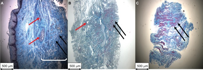
Figure 1.
Representative Masson trichome staining of 5 μm sections (magnification ×40) of a (A) full thickness vaginal biopsy of the anterior vaginal wall from a control patient used to measure vaginal wall thickness, (B) a cross-section of an anterior vaginal muscularis (AVM) strip from a control patient, and (C) a cross-section of an AVM strip from a woman with prolapse. (B) and (C) AVM strips were used to measure total and smooth muscle cross-sectional areas (mm2). Red arrows: blood vessel; black arrows: vaginal smooth muscle; white bracket: vaginal muscularis layer.

