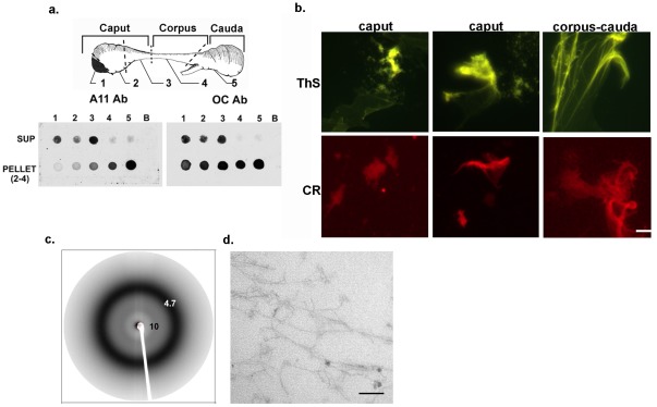
Figure 1. Amyloid structures in the mouse epididymal lumen.
a) Dot blot analysis using All and OC antibodies to detect the oligomeric and fibrillar amyloid structures, respectively, in supernatant and high speed pellet fractions isolated from the five regions of the mouse epididymis. b) Thioflavin S (ThS) and Congo Red (CR) bound to similar structures including a film-like material (third panel) in the high speed pellet isolated from the caput (regions 1–3) and corpus-cauda (regions 4–5) luminal fluid from the mouse epididymis. Bar, 5 µm. c) X-ray diffraction of a 250,000×g pellet (pellet 4) isolated from the corpus-cauda luminal fluid. d) Transmission electron microscopy of an Epon embedded high speed pellet isolated from the caput epididymal luminal fluid. Bar, 100 µm.

