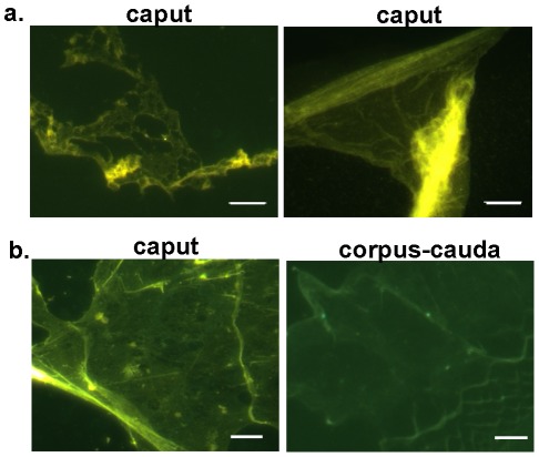
Figure 3. Thioflavin S staining of epididymal luminal fluid.
a) Caput luminal fluid was centrifuged to pellet spermatozoa and the resulting supernatant dried on a slide and stained with thioflavin S. Representative structures are shown. b) Luminal fluid was isolated after puncturing the caput and corpus-cauda epididymal tubules with a needle and allowing contents to disperse. After centrifugation to remove spermatozoa, the supernatant was dried on a slide and stained with thioflavin S. Bar, 5 µm.

