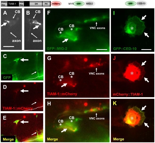
Figure 5. TIAM-1::mCherry is present in ectopic protrusions and growth cones and co-localizes with GFP::MIG-2 and GFP::CED-10.
The diagrams above the micrographs represent the fusion proteins produced by the transgenes used: TIAM-1 C-terminally tagged with mCherry; MIG-2 N-terminally tagged with GFP after the predicted myristoylation site; and CED-10 N-terminally tagged with GFP. For the NIH 3T3 studies (I–K), an N-terminally-tagged TIAM-1 was assayed. Panels are fluorescent micrographs of animals harboring transgenes that express TIAM-1::mCherry, GFP, or GFP::MIG-2. In (A–H), dorsal is up and anterior is to the left. (A and B) Micrographs of PDE neurons of animals carrying an osm-6p::tiam-1::mCherry transgene expressed in the PDE neurons. TIAM-1::mCherry accumulated in ectopic protrusions that occurred at a low frequency (large arrows). Small arrows point to the cell bodies (CB) and the axons, which are faint. (C–E) A VD growth cone (large arrow) in an early L2 animal 18–20 hours post-hatching. The growth cone was visualized using cytoplasmic unc-25p::gfp, and also showed TIAM-1::mCherry accumulation driven from the unc-25 promoter. The dashed line indicates the ventral nerve cord, and the small arrow points to a commissural motor axon that has completed commissural extension. The scale bar in (A) represents 5 µm for (A–E). (F–H) TIAM-1::mCherry and GFP::MIG-2 colocalized in ectopic protrusions. A ventral aspect of the ventral nerve cord is shown. (F) unc-25p::gfp::mig-2 expression in an ectopic protrusion from VD or DD motor neurons (arrow). Cell bodies are indicated (CB, asterisk), as is the ventral cord neuropil (VNC axons). (G) unc-25p::tiam-1::mCherry expression in the ectopic protrusion of the same VD/DD neuron. (H) Co-localization of GFP::MIG-2 and TIAM-1::mCherry in the ectopic protrusion from the VD/DD neuron. The scale bar in (F) represents 3 µm for (F–H). (I–K) CED-10::GFP and TIAM-1::mCherry expression in NIH 3T3 cells. (I) GFP::CED-10 accumulated in membranous regions around the nucleus (asterisk), as well as at the periphery of lamellipodial ruffles induced by GFP::CED-10 (arrows). (J–K) TIAM-1::mCherry co-localized with GFP::CED-10 at the periphery of CED-10::GFP-induced lamellipodial ruffles (arrows). mCherry::TIAM-1 also accumulated to a perinuclear region that was more widespread than GFP::CED-10 accumulation (asterisk). The scale bar in (I) represents 10 µm for (I–K).

