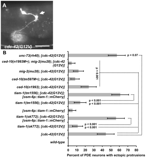
Figure 6. TIAM-1 acts cell autonomously downstream of CDC-42.
(A) Fluorescent micrograph of a PDE neuron from an animal with an integrated cdc-42(G12V) transgene expressed with the osm-6 promoter. An osm-6::gfp marker transgene was included to label the PDE cell body and processes. Dorsal is up and anterior is to the left. An arrow points to an ectopic lamellipodial protrusion. The scale bar in represents 5 µm. (B) A graph charting the percentage of PDE axons with ectopic lamellipodial and filopodial protrusions (X axis) in different genotypes (Y axis). Unless otherwise noted, all backgrounds are wild type. M+ indicates that the genotype had wild-type maternal gene function. [cdc-42(G12V)] indicates a transgene that harbors activated cdc-42(G12V) driven by the osm-6 promoter in the PDEs. [osm-6p::tiam-1(+)::mCherry] indicates a transgene that harbors the corrected tiam-1 cDNA yk730h9 fused in frame to mCherry and driven by the osm-6 promoter in the PDE neurons (see Methods and Figure S2). At least 100 PDE neurons were scored for each genotype, and p value significance was determined using Fisher's Exact analysis. Error bars represent 2× standard error of the proportion in both directions. Dashed lines indicate comparisons between genotypes not marked with a p value to those marked with p values.

