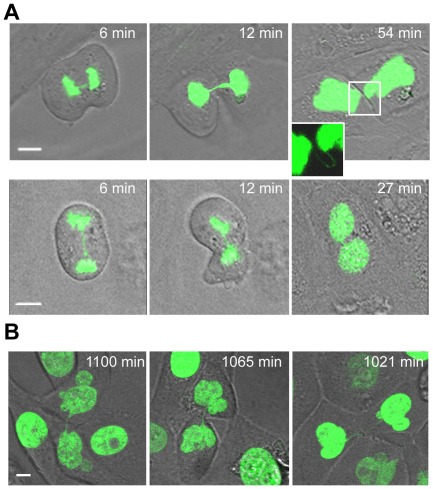
Figure 6. Unbroken anaphase bridges interfere with abscission in HMECs and HMEC-hTERT cells.
Non-transduced and hTERT-transduced HMECs were transfected with a H2B-GPF plasmid. (A) Time-lapse micrographies of non-transduced HMECs transiently expressing H2B-GFP. Time 0 was set as the last time point before anaphase onset. Upper images show progressively strengthened unbroken anaphase bridges that remain undisrupted connecting the two nuclei (see inset). Lower images show the formation of a binucleated cell where this process is coupled to the presence of an unbroken chromatin bridge. The scale bar represents 10 µm. (B) Three examples of HMEC-hTERT_BLEO expressing H2B-GFP with over strengthened chromatin bridges long time after anaphase onset. Scale bar represents 10 µm.

