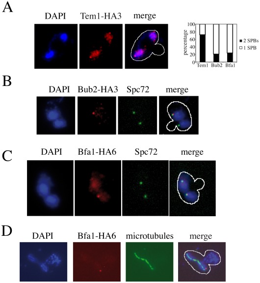
Figure 5. Asymmetric SPB localization of Bub2/Bfa1 in dma1Δ dma2Δ cla4-75 swe1Δ cells with mispositioned spindle.
A–C: Cells were grown in YEPD at 25°C and shifted to 37°C for 3 hours, followed by nuclear staining with DAPI and visualization of the indicated proteins and of the SPB component Spc72 by indirect immunofluorescence. Only micrographs of cells with mispositioned spindles are shown. A: dma1Δ dma2Δ cla4-75 swe1Δ cells (ySP7789) expressing HA-tagged Tem1 (Tem1-HA3). B: dma1Δ dma2Δ cla4-75 swe1Δ cells (ySP7771) expressing HA-tagged Bub2 (Bub2-HA3). C: dma1Δ dma2Δ cla4-75 swe1Δ cells (ySP7819) expressing HA-tagged Bfa1 (Bfa1-HA6). D: dma1Δ dma2Δ cla4-75 swe1Δ cells (ySP7819) expressing Bfa1-HA6 were grown in synthetic medium lacking uracil at 25°C, synchronized in G1 by alpha factor and released at 37°C, followed by nuclear staining with DAPI and visualization of Bfa1-HA6 and microtubules by indirect immunofluorescence. Micrographs were taken 90 minutes after release, when the percentage of mispositioned anaphase spindles was maximal.

