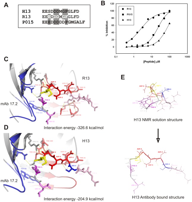
Figure 1. scFv C5 Epitope specificity.
A. Sequence aligment of T. cruzi R13 and P015 peptides with the mammalian counterpart H13 peptide. White letters correspond to residues necessary for antibody recognition, as identified by alanine scanning. Grey background corresponds to those residues conserved in the other two peptides. B. Inhibition of the interaction between scFv C5 and TcP2β protein by R13, P015 and H13 peptides, using surface plasmon analysis. The figure corresponds to one representative result out of 3 independent assays. C. Crystal structure of the complex mAb 17.2-R13 (PDB 3SGE). D. Model of the interaction of mAb 17.2 with the H13 peptide. The interaction energy is indicated on each case. E. NMR solution structure of H13 peptide (PDB 1S4J) in comparison with the antibody-bound peptide structure modeled.

