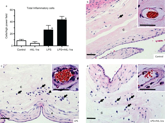
Figure 4.
Interleukin 1 (IL-1) signaling did not decrease lipopolysaccharide (LPS)-induced histologic chorioamnionitis in preterm fetal sheep. Assessments were made 2 days after intra-amniotic injections. a, Quantitation of inflammatory cells in the chorioamnion. b-d, Representative photomicrographs of chorioamnion stained with hematoxylin and eosin in fetuses exposed to (b) saline (controls), (c) intra-amniotic LPS, or (d) LPS + recombinant human IL-1 receptor antagonist (rhIL-1ra). The insets show a higher magnification of chorionic blood vessels. Exposure to LPS increased inflammatory cells (arrows) in the chorion, increased subepithelial mesenchyme thickness in the amnion, and induced vasculitis (chorion arteriolar thickness in inset). Inhibition of IL-1 signaling did not modify LPS-induced changes (n = 4 animals/group; A indicates amnion; C = chorion; Scale bar 50 μm; * P < .05 compared to controls).

