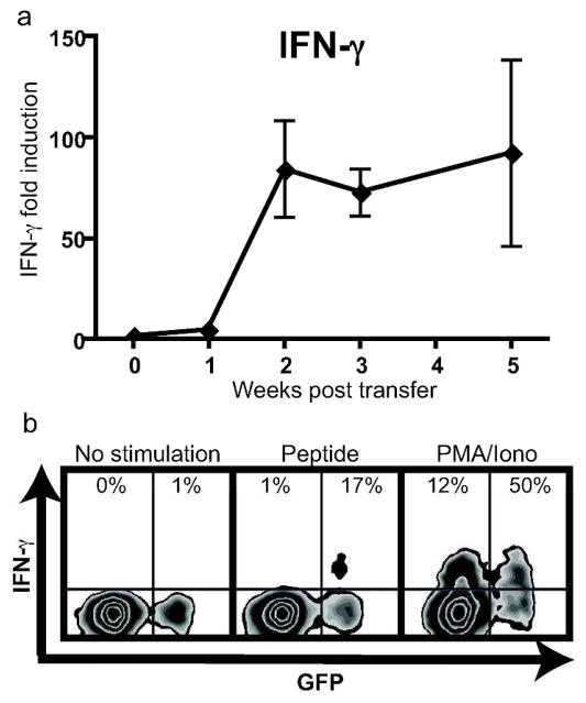
Figure 4. IFN-γ expression is induced in the skin during vitiligo and is produced by autoreactive CD8+ T cells.
(a) qRT-PCR for IFN-γ was performed on ear skin of mice at the indicated times (error bars = mean +/− SEM, n=3 mice per time point). (b) Single-cell suspensions were isolated from skin-draining lymph nodes 7 weeks after vitiligo induction and either left unstimulated, or stimulated with PMEL peptide or PMA and ionomycin as indicated. Following stimulation, cells were stained for IFN-γ, and flow cytometry revealed IFN-γ expression in transferred melanocyte-specific CD8+ T cells (GFP+) and endogenous CD8+ T cells (GFP−). Cells shown are gated on the total CD8+ population, and the percentage shown is of either the total GFP− or GFP+ gate. A representative result is shown.

