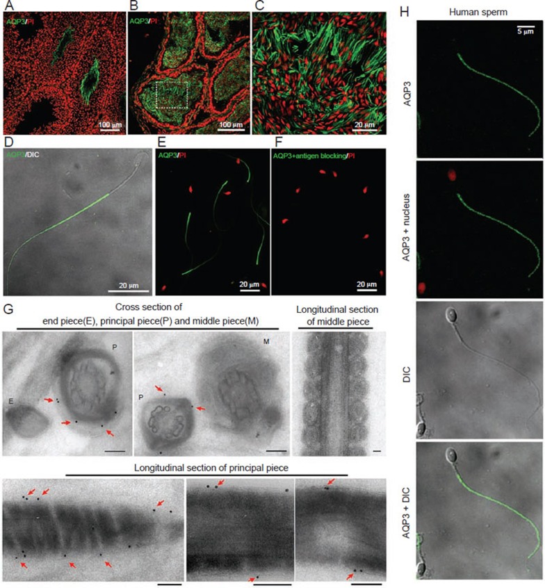
Figure 1.
AQP3 is expressed in mouse sperm. (A-D) Immunofluorescence staining of AQP3 in mouse testis (A), cauda epididymis (B, C) and isolated sperm (D) from cauda epididymis. Note the intensive green signal at principal piece of sperm tail. (E, F) AQP3 antibody staining in the absence (E) or presence (F) of competing immunogen. Nucleus was counter stained by propidium iodide (PI). (G) Immunogold-labeled electron microscopic detection shows that gold particles are stained at plasma membrane of the principal piece (indicated by arrows). Scale bars: 0.2 μm. (H) Immunofluorescence detection of AQP3 in human sperm.

