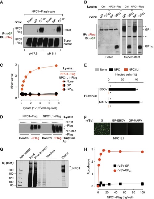
Figure 3.
NPC1 binds specifically and directly to a cleaved form of the Ebola virus glycoprotein. (A) Co-immunoprecipitation (co-IP) of NPC1 by EBOV GP. Magnetic beads coated with GP-specific monoclonal antibody KZ52 were incubated with detergent extracts containing no virus (None), uncleaved rVSV-GP, or cleaved rVSV-GPCL. The resulting control or glycoprotein-decorated beads were mixed with cell lysates containing human NPC1–Flag at pH 7.5 or 5.1 and 4°C. Beads were then retrieved and NPC1–Flag in the immune pellets and supernatants was detected by IB with an anti-Flag antibody. Pellets and supernatants were resolved on separate gels but exposed simultaneously to the same piece of film. (B) Reciprocal co-IP of GP by NPC1. Cell lysates lacking (Ctrl) or containing NPC1–Flag were incubated with anti-Flag antibody-coated magnetic beads. The resulting control or NPC1-decorated beads were mixed with detergent extracts of rVSV-GP or rVSV-GPCL at pH 7.5 and 4°C. Beads were then retrieved and GP or GPCL in the immune pellets and supernatants was detected by IB with an anti-GP antiserum. Pellets and supernatants were resolved on separate gels but exposed simultaneously to the same piece of film. Asterisks indicate bands detected non-specifically by the antiserum. (C) GPCL captures NPC1 but not NPC1-like1 (NPC1L1) in an ELISA. Plates coated with rVSV-GP or rVSV-GPCL were incubated with cell extracts containing NPC1–Flag or NPC1L1–Flag, and bound Flag-tagged proteins were detected with an anti-Flag antibody. Results (n=3) are representative of at least four independent experiments. (D) Cell extracts used in (C) were incubated with plates coated with an anti-Flag antibody or an isotype-matched control, and captured proteins were eluted and detected by IB with the anti-Flag antibody. Samples were resolved on the same gel. (E, F) NPC1L1 cannot support filovirus entry and infection. CT43 cells expressing NPC1L1–Flag were exposed to wild-type EBOV or MARV (E) or to recombinant VSVs (F), and infected cells were visualized and enumerated by fluorescence microscopy. Asterisks in (E) indicate values below the limit of detection. Scale bar, 20 μm. (G, H) GPCL but not GP captures affinity-purified NPC1–Flag in an ELISA. (G) NPC1–Flag was purified from CT43 CHO cell lysates by Flag affinity chromatography and visualized by SDS–PAGE and staining with Krypton infrared protein stain. Samples were resolved on the same gel. (H) ELISA plates coated with rVSV-GP or rVSV-GPCL were incubated with NPC1–Flag purified in (G), and bound Flag-tagged proteins were detected with an anti-Flag antibody. Results (n=3) are representative of at least four independent experiments. Error bars indicate s.d. Figure source data can be found in Supplementary data.

