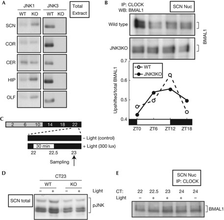
Figure 5.
BMAL1 phosphorylation in the mouse suprachiasmatic nucleus (SCN). (A) The total extracts of indicated brain regions were prepared at ZT6 from wild-type mice (WT) or Jnk3-deficient mice (KO). Equal protein amounts of the samples were subjected to immunoblot analysis. CER, cerebellum; COR, cortex; HIP, hippocampus; OLF, olfactory bulb. (B) The SCN nuclear extracts were prepared at ZT0, 6, 12 and 18 from 15 mice (for each time point) of WT and KO mice. They were immunoprecipitated (IP) with anti-CLOCK antibody and subjected to immunoblot analysis with anti-BMAL1 antibody. The relative level of the upshifted band was plotted at the bottom. (C) Time schedules for SCN sampling on the first day of constant darkness (DD). (D) Light-induced change of JNK phosphorylation level in the SCN of WT and KO mice was investigated after exposure to 30-min light pulse at circadian time (CT)22. Total extract was prepared from the SCN punch of WT and KO mice, and subjected to immunoblot analysis with anti-phospho-JNK antibody. (E) Light-induced change of BMAL1 phosphorylation level in the SCN of WT mice was investigated after exposure to 30-min light pulse at CT22. JNK, c-Jun N-terminal kinase; WB, western blot.

