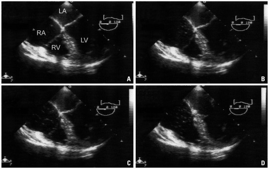
Fig. 1.
Carbon dioxide emboli detected by transesophageal echocardiography in a patient undergoing total laparoscopic hysterectomy; mid-esophageal four chamber view. (A) A single gas bubble in the right atrium (RA), right ventricle (RV), and right ventricle outflow tract (RVOT) (grade I). (B) gas bubbles filling less than half the diameter of RA, RV, and RVOT (grade II). (C) gas bubbles filling more than half the diameter of RA, RV, and RVOT (grade III). (D) gas bubbles completely filling the diameter of RA, RV, and RVOT (grade IV). Permission from Kim, et al.13

