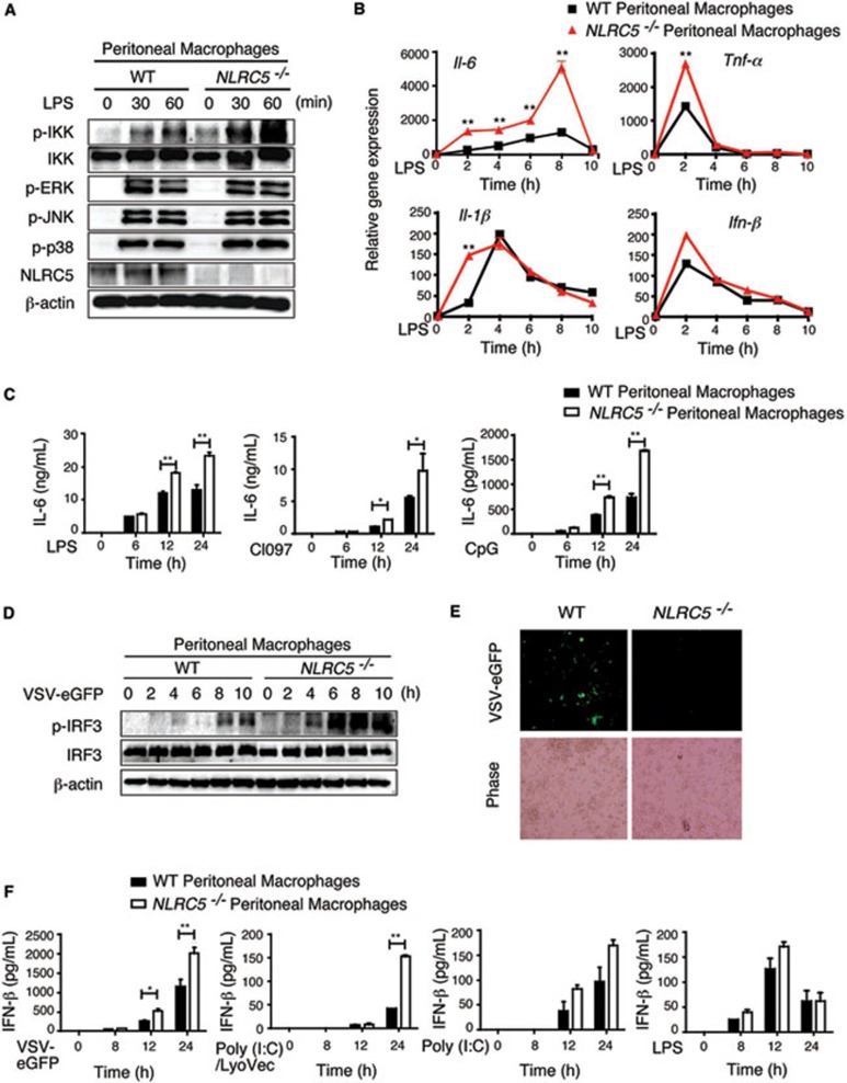
Figure 5.
NLRC5 deletion enhances both NF-κB and type I IFN signaling, as well as cytokine production in peritoneal macrophages. (A, B) NLRC5−/− and WT peritoneal macrophages were incubated with LPS (100 ng/ml) for the indicated times. The cell lysates were harvested for immunoblotting with the indicated antibodies (A). The RNA was collected for real-time PCR analysis (B). (C) NLRC5−/− and WT peritoneal macrophages were incubated with LPS, CL-097 or CpG for the indicated times, and the culture supernatants were used for ELISA analysis to measure the production of IL-6. (D) NLRC5−/− and WT peritoneal macrophages were infected with VSV-eGFP for the indicated times. Cell lysates were harvested to detect IRF3 phosphorylation by immunoblotting. (E) Peritoneal macrophages were infected with VSV-eGFP (MOI = 10) for 24 h, then viral infection was analyzed by fluorescence microscopy (with phase contrast as a control). (F) NLRC5−/− and WT peritoneal macrophages were treated with VSV-eGFP, poly(I:C)/LyoVec, poly(I:C) or LPS for the indicated times, and the culture supernatants were used for ELISA analysis to measure the production of IFN-β. B, C and F are plotted as means ± SD. Results are representative of three independent experiments. *P < 0.05, **P < 0.01, versus controls.

