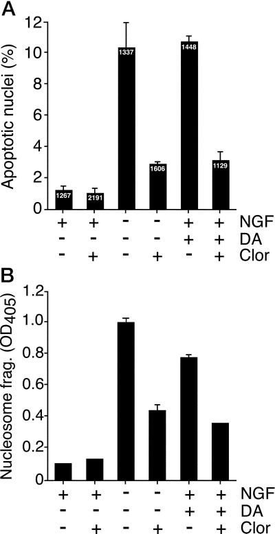
Figure 6.
Effect of MAO inhibition on dopamine and NGF withdrawal-induced apoptosis. Neuronal PC12 cells were incubated (24 h) in the presence and absence of NGF, or in the presence of NGF plus 300 μM dopamine (DA). The effect of the addition of 0.1 μM clorgyline (Clor) was examined. (A) The cells were fixed, stained with 4,6-diamidino-2-phenylindole, and inspected by fluorescence microscopy. Cells with condensed chromatin and/or fragmented nuclei were scored as apoptotic. The number of cells examined for each treatment group is shown (Inset). The percentage of apoptotic cells (mean ± SEM; n = 3) is presented. (B) Apoptosis was examined by measurement of nucleosomal DNA fragmentation (mean ± SD; n = 3). Similar data were obtained in three independent experiments.

