Figure 2.
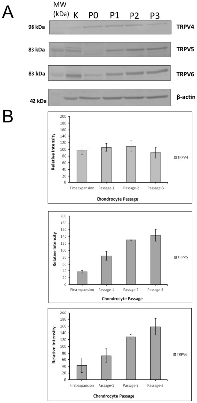
(A) Western blot analysis of total chondrocytes lysate (25 μg/lane) from first expansion (P0) and serial passages (P) 1–3. Kidney was used as a positive control. Western blotting confirmed the expression of TRPV4, TRPV5 and TRPV6 proteins in different passages. B. Image analysis of western blots. No significant change in expression profile was observed in TRPV4 among different passages. TRPV5 and TRPV6 expression was significantly upregulated with time and passage in culture (P < 0.05).

