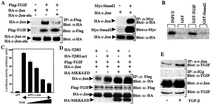
Figure 4.
Association of c-Jun with TGIF. (A) Cell lysates from transiently transfected COS-7 cells were subjected to immunoprecipitation with anti-Flag or anti-Myc antibodies and then immunoblotted by using anti-HA that recognizes HA-c-Jun or HA-c-Jun-ala. (B) In vitro interaction of c-Jun with TGIF or Smad2 was examined by incubating full-length [35S]methionine-labeled c-Jun produced by in vitro transcription/translation with Sepharose-bound bacterially expressed GST-TGIF, GST-Smad2, or GST. Bound material was visualized by SDS and autoradiography. Ponceau staining of the membrane showed that similar amounts of GST, GST-Smad2, and GST-TGIF were used in this assay (data not shown). (C) HepG2 cells were cotransfected with AP1-Lux together with c-Jun and increasing amounts of TGIF. After 48 h, luciferase activity was determined and normalized to β-galactosidase activity. (D) COS-7 cells were transfected with wild-type Flag-TGIF and HA-c-Jun, together with HA-TβRI or HA-TβRI.act, either in the absence or the presence of MKK4.ED. Cell lysates were subjected to anti-Flag immunoprecipitation and then immunoblotted with anti-HA antibody. (E) Proteins were precipitated from Mv1Lu and from Mv1Lu cells treated with TGF-β for 1 h by using an anti-rabbit polyclonal antibody specific for c-Jun (Oncogene; Top) or normal rabbit antiserum (Middle). Precipitated proteins were analyzed by Western blotting with a goat antibody specific for TGIF (Santa Cruz). For comparison, a portion of cell lysates was probed with anti-c-Jun or anti-TGIF (Bottom) antibodies.

