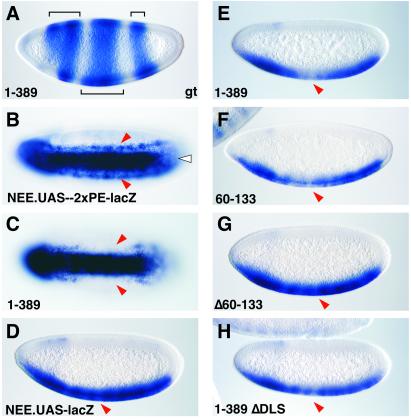
Figure 4.
Activities of Gal4-Giant fusion genes. Transgenic embryos contain either the NEE.UAS-2xPE-lacZ or NEE.UAS-lacZ reporter gene and different Krüppel-Gal4-Giant expression vectors. Embryos were hybridized with either a giant antisense RNA probe (A) or a lacZ probe (B–H). (A) giant staining pattern in a transgenic embryo that contains the Krüppel-Gal4-Giant (1) fusion gene. Staining is detected in anterior and posterior regions (Upper, brackets), which correspond to the endogenous pattern. The Krüppel transgene directs strong expression in central regions (Lower, bracket). (B) The NEE.UAS-2xPE-lacZ reporter gene in a wild-type embryo lacking any of the Krüppel-Gal4-Giant expression vectors. Staining is detected in the ventral mesoderm (white arrowhead) and in lateral lines in the neurogenic ectoderm (red arrowheads). (C) Same as B, except that the embryo also expresses the Gal4-Giant (1) fusion protein. The lateral staining pattern directed by the modified NEE is repressed in central regions (arrowheads). In contrast, staining directed by the twist enhancer (2xPE) is unaffected. (D) The NEE.UAS-lacZ reporter gene in a wild-type embryo lacking Krüppel-Gal4-Giant expression vectors. The lacZ reporter gene exhibits uniform expression in ventral regions. There is no gap in the center (arrowhead). (E) Same as D, except that the embryo also expresses the Gal4-Giant (1) fusion protein. There is a gap in central regions (arrowhead). (F) Same as E, except that the embryo expresses a Gal4-Giant fusion protein containing the minimal Giant repression domain (amino acid residues 60–133). There is a gap in the staining pattern in central regions (arrowhead). (G) Same as D, except that the embryo also expresses a mutant version of the full-length Gal4-Giant fusion protein with an internal deletion in the minimal repression domain (Δ60–133). There is no significant repression of lacZ staining in central regions (arrowhead). (H) Same as D, except that the embryo also expresses a mutant form of the Gal4-Giant fusion protein containing alanine substitutions in the VLDLS motif (see Fig. 3).

