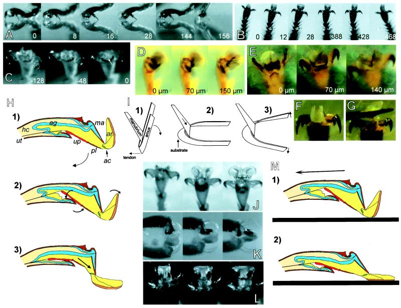
Figure 2.
(A–C) High-speed recordings of steps taken upside down on a glass plate (numbers indicate time in milliseconds). (A) A. mellifera, lateral view. (B) A. mellifera, ventral view. (C) O. smaragdina, arolium deflation before detachment (arrow indicates point of detachment). (D and E) O. smaragdina, experimental pull on the unguitractor tendon; numbers indicate the amplitude of the tendon pull (“0” is defined as the tendon-pull amplitude where the first pretarsus movement was visible). (D) Pretarsus lateral view. (E) Ventral view, arolium surface focused. (F) A. mellifera, arolium at maximal pull of the tendon (200 μm). (G) Same as F, arolium spread laterally by application of upward pressure to the planta with an insect pin. (H) Model of arolium extension caused by the contraction of the claw-flexor muscle. For abbreviations see legend for Fig. 1. (I) Model of the interaction between the two arolium sclerites, arcus and manubrium. (J–L) Passive extension of arolium in contact with a glass surface. (J) O. smaragdina, pull of severed leg in the direction toward the body. (K and L) Pull of legs of freshly killed A. mellifera toward the body, lateral and frontal view of arolium, respectively. (M) Model of passive arolium extension caused by substratum contact and horizontal pull of the leg.

