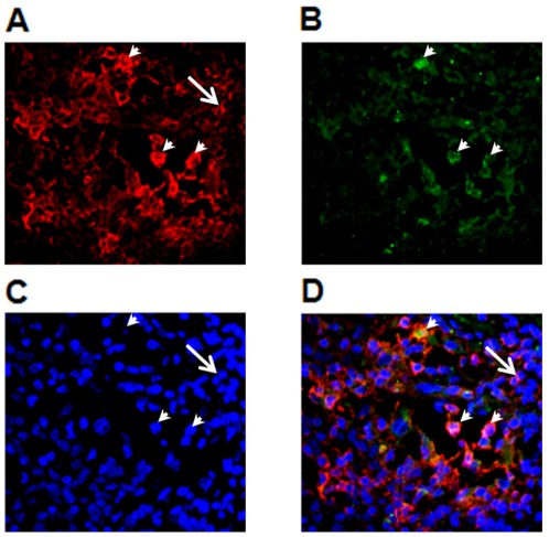
Figure 8. CTLA-4 is expressed by infiltrating T lymphocytes in the heart of chronic Chagas disease patients.
Double immunofluorescence staining with CD3 and CTLA-4 antibodies was performed as described in Material and Methods. From total CD3-expressing T cells present in the inflammatory infiltrate (A) a small proportion showed CTLA-4 expression (B). Nuclei staining with DAPI. The arrowheads point the nuclei of CTLA4+ cells (C). Composite of figures A, B and C showing the double stained cells (arrowheads) and a CD3+CTLA-4− single stained cell (large arrow)(D). Original Magnification 400×.

