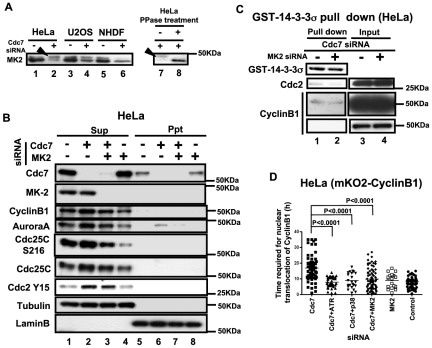
Figure 5. MK2 is activated in Cdc7-depleted HeLa cells and is required for cytoplasmic accumulation of CyclinB1.
(A) HeLa (lanes 1, 2, 7 and 8), U2OS (lanes 3 and 4) and NHDF (lanes 5 and 6) cells were treated with control or Cdc7 siRNA and the whole cell extracts were run on a phosgel and analyzed by western blotting. Lanes 7 and 8, extracts from Cdc7 siRNA-treated HeLa cells were non-treated (−) or treated with λ-phosphatase (+). Arrowheads indicate the phosphorylated MK2 band. (B) HeLa cells were treated with control siRNA (lanes 1 and 5), Cdc7 siRNA (lanes 2 and 6), Cdc7 and MK2 siRNAs (lanes 3 and 7) and MK2 siRNA (lanes 4 and 8) for 48 hrs, and CSK-soluble (lanes 1–4; Sup) and -insoluble (lanes 5–8; Ppt) proteins were analyzed by western blotting. Tubulin and LaminB are shown as loading controls. (C) Glutathion Sepharose 4B beads carrying GST-14-3-3σ protein was incubated for 1 hr at 4°C with CSK-soluble extracts of HeLa cells treated with siRNA, as shown. Bound proteins were examined by Western blotting. “Input” represents only the extracts without added GST-14-3-3σ protein. Cdc7 and MK2 co-depletion reduced the binding between 14-3-3σ and Cdc2/CyclinB1. (D) HeLa cells expressing mKO2-CyclinB1 were treated with indicated siRNAs. The time (hr) between the first appearance of cytosolic mKO2-CyclinB1 signal and its translocation into the nucleus was measured in the time lapse images. Co-depletion of MK2, p38 (upstream kinase of MK2) or ATR reduced cytoplasmic retention of CyclinB1 (G2 elongation) was observed in Cdc7-depleted HeLa cells. The P-values of the two-tailed unpaired t-test were calculated by Prism software. Cdc7-D siRNA was used in all the experiments.

