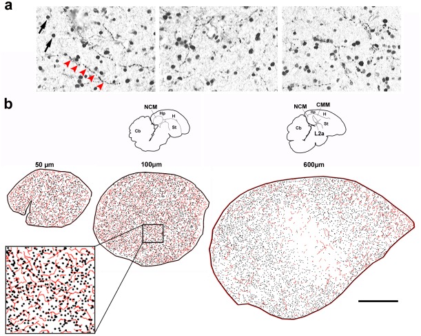
Figure 1. Noradrenergic innervation and sensory-activated neurons in NCM.
a) Representative images of double-immunostaining for ZENK (nuclear) and DBH (fibers) in parasaggital brain sections of song-stimulated zebra finches; red arrowheads depict a DBH-positive fiber. b) Camera lucida drawings from serial brain sections depicting DBH-positive fibers (red traces) and ZENK-expressing neurons (black dots) at different medial to lateral NCM levels (in µm from the midline); NCM location is indicated by the small brain diagrams on top and the inset at the bottom left shows a detailed view of the tracings, at about half the magnification as the panels in a; the brain section containing the most medial NCM (50 µm from the midline) is often incomplete and therefore is not represented here. Abbreviations: Cb, cerebellum; CMM, caudomedial mesopallium; Hp, hippocampus; H, hyperpallium; L2a, subfield L2a of field L; NCM, caudomedial nidopallium; St, striatum. Scale bar, 500 µm.

