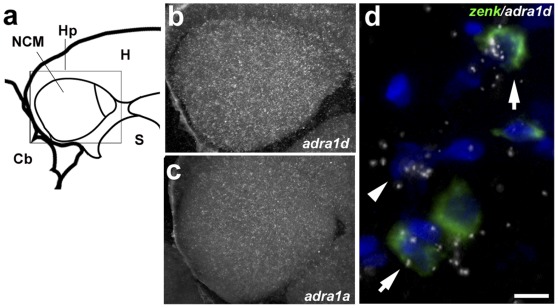
Figure 6. Expression of α-adrenergic receptors in NCM.
a) Camera lucida drawing of a parasagittal brain section containing NCM and CMM (about 100 µm from the midline), indicating the position of the photomicrographs in b and c. b–c) Dark-field view of emulsion autoradiograms of brain sections hybridized with radioactively-labeled ADRA1d and 1a riboprobes, respectively; shown is an area corresponding to the rectangle in a. d) High-magnification view of double in situ hybridization for ADRA1d (emulsion grains) and zenk (green fluorescence); arrows point to double-labeled cells and arrowhead indicates a single-labeled ADRA1d-positive cell.

