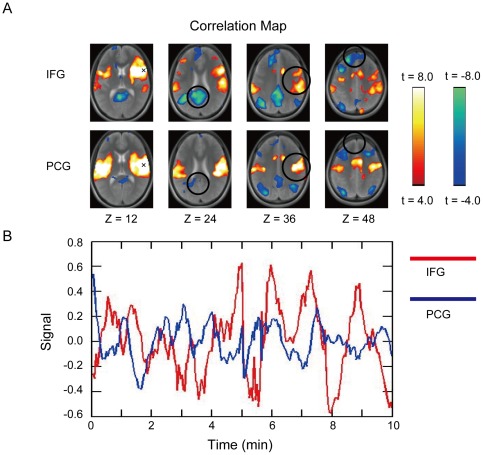
Figure 2. Different spatial patterns in whole-brain correaltion maps and the signal timecourses between the 2 seeds as close as 2 cm.
A) Whole-brain correlation maps that exhibited distinct spatial patterns when seeds were placed around the right pIFC about 2 cm apart, at (54, 12, 12) and (54, −8, 12) [51]. A seed is indicated by “x”. IFG: inferior frontal gyrus, PCG: precentral gyrus. B) fMRI signals in the two seed points in one representative subject that exhibited dissimilar timecourses. The correlation between the timecourses was utilized in the present study for boundary mapping.

