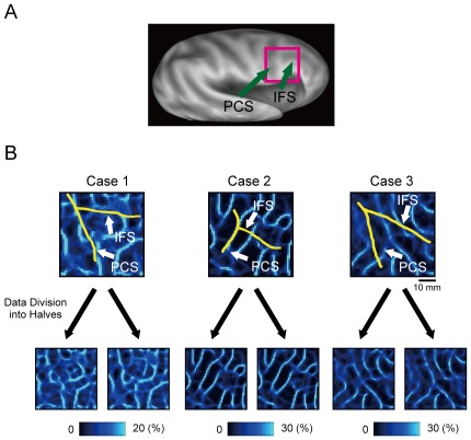
Figure 3. The probabilistic boundary maps around the right pIFC generated by the standard analysis with whole-brain calculation of correlation.
A) An inflated brain and the region of interest, the right pIFC. PCS: precentral sulcus, IFS: inferior frontal sulcus. B) Reproducibility of boundary patterns based on the standard mapping method in three individual subjects when data set was divided into two halves (left panels: odd runs, right panels: even runs). Yellow curves indicate the approximate location of the fundus of the PCS or IFS.

