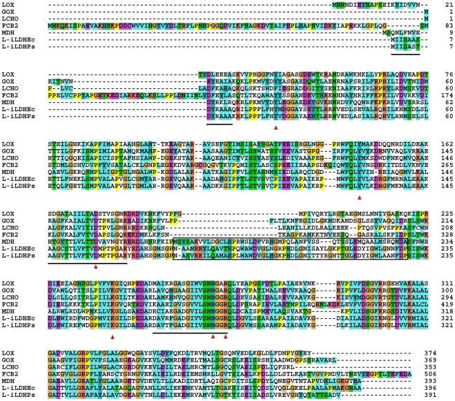
Figure 3. Sequence alignment of l-iLDH from P. stutzeri SDM with other FMN-dependent α-hydroxy acid oxidases.
LOX, l-lactate oxidase in A. viridans [19]; LCHO, long-chain α-hydroxy acid oxidase in rat kidney [20]; MDH, l-mandelate dehydrogenase in P. putida [21]; GOX, glycolate oxidase in S. oleracea [22]; FCB2, flavocytochrome b2 (l-iLDH) in S. cerevisiae [18]; l-iLDHPs, l-iLDH in P. stutzeri SDM; l-iLDHEc, l-iLDH in E. coli [9]. Red arrows indicate the highly conserved residues important for FMN binding and enzymatic catalysis across this class of enzymes. The boxed segments in l-mandelate dehydrogenase represent the internal sequences that were implicated in membrane association. The lined segments in l-iLDH in P. stutzeri SDM represent the peptides identified through Edman degradation analysis and ESI-MS/MS. The sequences were aligned with the program CLUSTAL X [23]. Parameters and color scheme were set as default.

