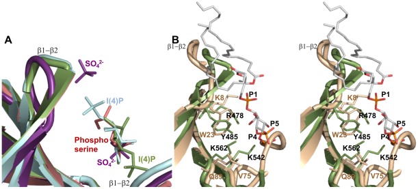
Figure 3. Superposition of Slm1-PH domain structures.
(A) The different ligands (sulfate (violet), phosphoserine (salmon), Ins(4)P (green) and also the turned-over Ins(4)P (light blue)) are occupying the same non-canonical binding pocket, confirming the readily available large binding site of the Slm1-PH. (B) Stereo diagram of PtdIns(4,5)P2 (atom colors) modeled in the non-canonical binding site of Slm1-PH (green) and overlaid onto the β-spectrin PH domain (wheat color). This shows the conservation of side chains that contact the ligands.

