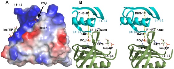
Figure 4. Surface charge distribution.
(A) The Slm1-PH provides a positively-charged cavity to interact with a negatively charged Ins(4)P molecule (in green). The back of the β1-β2 region (by rotating Slm1-PH by 180 degrees) shows a more positively charged region. In several structures of Slm1-PH determined in complex with ligand analogs, we built a phosphate group or full Ins(4)P at this additional positively charged site (see text for details). The Arg477 side chain that turns towards the canonical binding site is also shown in transparent stick format. (B) Stereo diagram of Slm1-PH (green) showing Arg477, Arg478, Lys480 and Lys483 on either side of the β1-β2 strands in the vicinity of the negatively charged residues of the β5-β6 loop of the neighboring molecule (cyan). Part of the β5-β6 loop is truncated for the clarity of the figure. The bound phosphate group is shown in stick representation at the back of the β1-β2 region towards the canonical binding site. We have modeled the natural ligand (DHS-1P in yellow) aligning with the phosphate position in this region. Ins(4)P is also shown in the non-canonical binding site.

