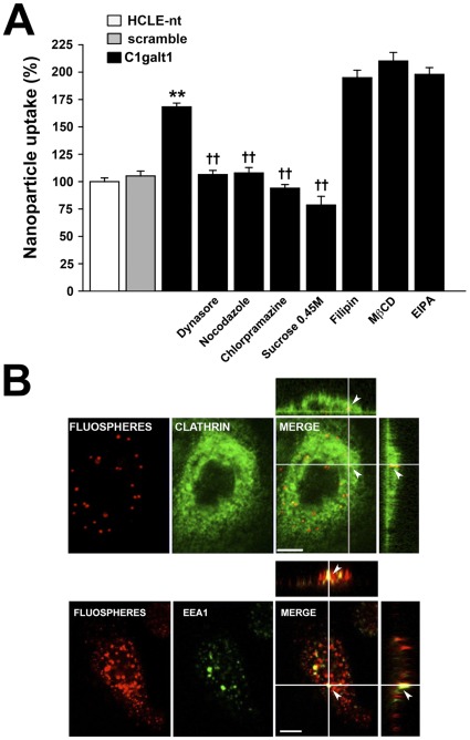
Figure 5. Clathrin-mediated endocytosis is a predominant pathway for nanoparticle internalization in C1galt1 shRNA cells.
(A) For fluorometry assays, corneal keratinocytes were pre-incubated at 37°C in the presence of inhibitors of endocytosis. After 30 min, FluoSpheres® suspensions (1010 nanoparticles/ml) were added to the cells in the continuous presence of inhibitors and incubated for 3 h at 37°C. Sucrose was directly added to the suspension in these experiments. Nanoparticle uptake was quantified by fluorometry as described in Materials and Methods. Values are normalized to percent total uptake in HCLE-nt cells. Data for each condition are reported as the mean of three wells in three independent experiments. **P<0.001 compared with scramble shRNA group and ††P<0.001 compared with the C1galt1 shRNA group with no inhibitors. (B) Confocal microscope images of C1galt1 shRNA cells. For colocalization experiments, keratinocytes were incubated with FluoSpheres®suspensions (red) for 3 h at 37°C. The cells were then fixed, permeabilized and stained for clathrin heavy chain or EEA1 (green). Examination of Z-stacked images shows colocalization of nanospheres with clathrin (Pearson's coefficient: 0.939) or EEA1 (Pearson's coefficient: 0.885) in the cytoplasm (arrowheads). Scale bar, 10 µm.

