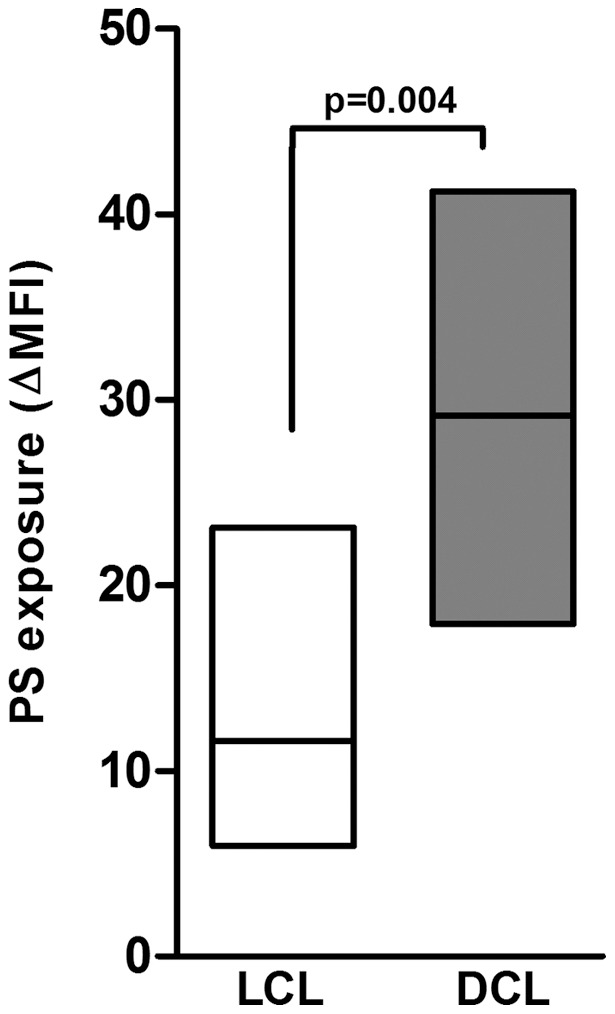
Figure 1. PS exposure on the L. amazonensis amastigotes surface.

Thioglycollate-induced peritoneal macrophages derived from F1 (BALB/C X C57BL/6) mice were infected with different isolates obtained from patients with LCL (BA69, BA73, BA115, BA 125, and M2269) (□ ) and DCL (BA106, BA276, BA336, BA700, and BA760) (▪ ) at a 3∶1 parasite-to-cell ratio. After 24 h of infection, amastigotes were purified for PS exposure analysis by flow-cytometry, as described in Methods. One representative experiment of at least five independent repeats is shown. Boxes represent median values and interquartile interval from different isolates mentioned above. Differences were checked using Unpaired t test.
