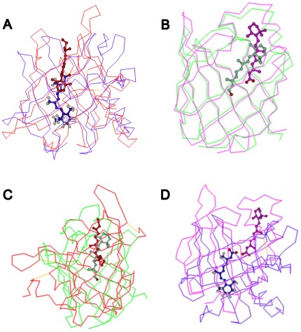
Figure 1. Comparison of retinoid binding in their transport proteins.
In the same protein family and subcellular location, retinol and retinoic acid have the same binding orientation (A, B), while in different protein families and subcellular locations, the binding orientations of the ligands are completely different (C, D). (a) In both of the extracellular proteins (RBP, ERABP), the β-ionone ring of the ligand is positioned in the center of the barrel with the isoprene tail extending along the barrel axis pointing toward the solvent. (b) The orientation of the ligand is, therefore, opposite to that in the corresponding intracellular retinoid-binding proteins (CRBPs and CRABPs). The red, blue, green and pink lines indicate RBP (PDB code: 1brp), ERABP (PDB code: 1epb), CRBP (PDB code: 1crb), and CRABP (PDB code: 1cbs) protein structures, respectively. The colors representing retinol and retinoic acid correspond to different transport protein colors.

