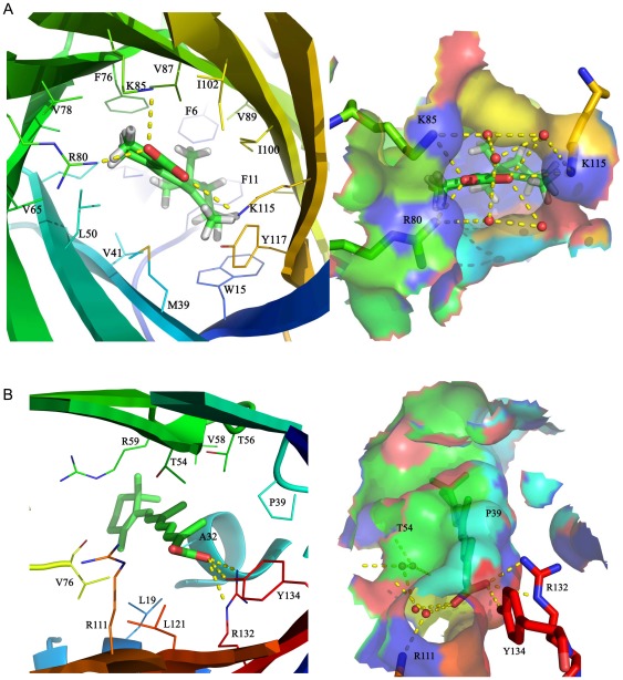
Figure 3. The retinoic acid-binding cavity in ERABP and CRABP.
The retinoic acid binding sites are displayed and colored as in Figure 2. (A) In ERABP, three positively charged amino acids (Arg80, Lys85 and Lys115) along with the retinoic acid carboxylate form a network of three ion pairs at the entrance end of the amphipathic binding site. Additional polar side chains and water molecules that participate in the network are included. (B) The carboxylate of the ligand interacts with a trio of residues (Arg132, Tyr134 and Arg111) in CRABP.

