Fig. 1.
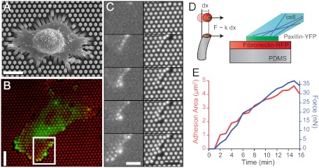
Cell adhesion and traction forces developed by REF52 fibroblasts expressing YFP-paxillin on micropillar substrates. (A) Scanning electron micrograph image of a typical REF52 cell on a micropillar substrate. (Scale bar, 15 µm.) (B) Epifluorescent image of a single cell deforming the micropillar substrate (here of spring constant k = 34 nN/μm). Micropillars are labeled by Cy3-fibronectin (red), and YFP-paxillin-rich patches are in green. (Scale bar, 15 µm.) (C) Sequential images of the insert area of B showing the dynamics of FA growth and micropillar displacements. (Scale bar, 10 µm.) (D) Schematic representation of the experimental setup showing the formation of FAs on the top of a PDMS micropillar. (E) Typical example of the formation of an FA area (red) and the buildup of force (blue) as a function of time (on a substrate of 34 nN/μm).

