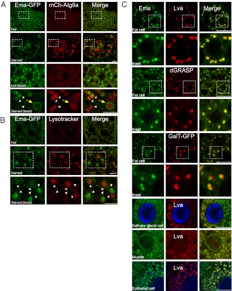
Fig. 5.
Ema localizes to the Golgi complex and moves to autophagosomes under starvation conditions. (A and B) Localization of Ema protein to autophagosomes during autophagy. (A) Representative single confocal sections of fed and starved fat body cells expressing Ema-GFP (green) and mCherry-Atg8a (red). Insets show autophagosomes with (white arrows) or without (arrowhead) Ema-GFP. Note that Ema-GFP localizes at the perimeter of autophagosomes (yellow arrows). (B) Representative single confocal sections of fed and starved fat body cells expressing Ema-GFP (green) and stained with LysoTracker DND-99 (red). Insets show the lack of colocalization of Ema-GFP (arrowheads) and LysoTracker structures (arrows). (C) Ema protein localizes to the Golgi complex. Representative single confocal sections of fat body cells, salivary gland cells, muscles, and epithelial cells expressing Ema protein tagged with either GFP (green) or mCherry(red) and labeled for the Golgi proteins Lva, dGRASP-GFP, or GalT-GFP. (Scale bars, 10 μm.)

