Fig. 4.
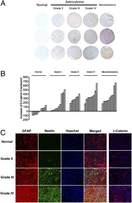
Changes associated with reactive astrocytes in astrocytomas of varying grade. (A) Tissue microarray analysis of β-catenin expression in astrocytomas of varying grade demonstrating a positive correlation of expression of β-catenin with the grade of the astrocytoma. (Magnification: 40×.) (B) Semiquantitative analysis of expression of β-catenin in samples of normal brain and tumors (individual bars) also demonstrating a positive correlation between expression of β-catenin and the grade of the astrocytoma. (C) Immunofluorescence staining for GFAP (red) and Nestin (green) demonstrating an increase in expression of these reactive astrocyte biomarkers in astrocytomas of increasing grade. In all astrocytomas, the pattern of expression of these markers appears heterogeneous and dysregulated. Far right column: Immunofluorescence staining for β-catenin (red) demonstrating increased β-catenin in astrocytomas of increasing grade. (Magnification: 200×.)

